- Share
- Share on Facebook
- Share on LinkedIn
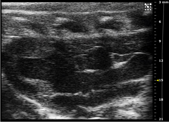
Ultrasound imaging is based on the reflection of ultrasonic waves. A transducer emits ultrasound waves that interact with organs, fluids, or internal structures. Depending on the density and nature of the tissues encountered, a portion of the waves is reflected back to the transducer. By analyzing the time and intensity of the returning waves, real-time images are reconstructed. Areas of differing density (bone, fluids, soft tissues) appear in varying shades of gray or color.
Doppler ultrasound, operating on the same principle, detects and quantifies the velocity of moving structures, enabling the study of blood flow. The frequency of the emitted waves determines both the resolution of the reconstructed image and the depth of exploration. Different transducers are therefore tailored to specific organs or models.
The GAIA platform is equipped with a state-of-the-art ultrasound imaging system (Vevo F2, FUJIFILM VisualSonics), offering high-resolution imaging that combines ultra-high and low frequencies. This system enables examinations on rodent models such as the assessment of cardiac ejection fraction in TM or mode B, vascular Doppler, strain rate, or stress ultrasound (coronary reserve), with in addition thanks to the associated manipulation platform, the possibility of monitoring respiratory and heart rate (ECG) and ensuring their synchronization with images ("gating"); but also tumor detection (3D engine available) or the characterization of tumor vasculature (contrast ultrasound).
Available Transducers:
-
22 MHz UHFx Series Transducer (Frequency range: 22–10 MHz): Ideal for cardiovascular and abdominal imaging in rats (>400g) or rabbits.
- 46 MHz UHFx Series Transducer (Frequency range: 46–20 MHz): Suited for cardiovascular imaging in mice, abdominal imaging in rats, ophthalmic imaging in rabbits, and vascular imaging (mice, rats, rabbits).
- Share
- Share on Facebook
- Share on LinkedIn