- Share
- Share on Facebook
- Share on LinkedIn
Background
Cardiovascular diseases remain the leading cause of death worldwide, with ischemic heart disease (IHD) being the primary contributor to cardiovascular mortality. Ischemic heart disease is caused by micro- and macrovascular coronary disease.
At the microvascular level, IHD is caused by early, diffuse and persistent damage to the coronary microcirculation, a process that remains impossible to assess non-invasively in routine clinical practice, despite its underlying mechanisms being relatively well understood.
At the macrovascular level, acute coronary syndromes, the main cause of myocardial infarction, typically result from the sudden rupture of so-called vulnerable atherosclerotic plaques. These plaques are strongly associated with inflammatory processes, making early detection a major clinical challenge.
Beyond atherosclerosis, chronic inflammation, marked by the recruitment of immune cells within the microvasculature, plays a key role in a wide range of diseases affecting multiple organs.
Objectives
The cardiovascular research theme at the LRB focuses on developing early diagnostic and prognostic tools, as well as methods to monitor therapeutic response. These rely on existing radiopharmaceuticals and the development of new tracers, used in nuclear imaging to investigate coronary microvascular dysfunction, the vulnerability of atherosclerotic plaques, and chronic inflammation in conditions such as spondyloarthritis and myocarditis.
In parallel, the LRB’s cardiologists are also involved in designing medical devices and developing advanced image analysis tools to improve patient care.
Coronary Microvascular Dysfonction
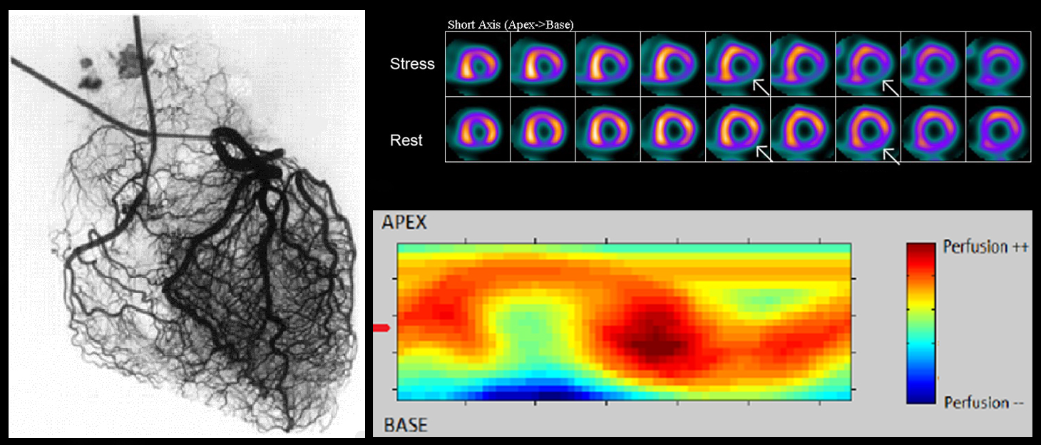
Recent studies have highlighted the significant impact of coronary microvascular dysfunction (CMD) on cardiovascular mortality. These advances have been made possible by the development of invasive tools for assessing coronary physiology. However, such methods remain limited to highly selected patient populations, and no non-invasive approach is currently available to assess the coronary microcirculation in vivo in humans, despite its high diagnostic and prognostic value.
The LRB focuses on the non-invasive assessment of CMD using myocardial perfusion imaging (MPI) with SPECT (Single Photon Emission Computed Tomography). MPI is a widely used, non-invasive technique for diagnosing and stratifying cardiovascular risk through nuclear stress testing and is routinely performed in millions of patients worldwide.
Working with patient cohorts and registries from the Grenoble-Alpes University Hospital (CHU) and in collaboration with both local and international experts, the LRB is also developing AI tools to enhance the diagnostic and prognostic performance of SPECT myocardial perfusion imaging in the evaluation of coronary microvascular dysfunction.
Atherosclerosis
Atherosclerosis Imaging
Atherosclerotic plaques are lesions that develop within the arterial wall. When they form in the coronary arteries that supply the heart, they can lead to the development of occlusive thrombi, which in turn causes myocardial infarction. While stenotic plaques, those that significantly narrow the vessel lumen, can be detected through coronary angiography, this technique does not reliably identify vulnerable plaques, which typically grow eccentrically and only mildly affect the lumen.
Yet these vulnerable plaques are responsible for the majority of acute myocardial infarctions, and their early detection remains a major unmet clinical need.
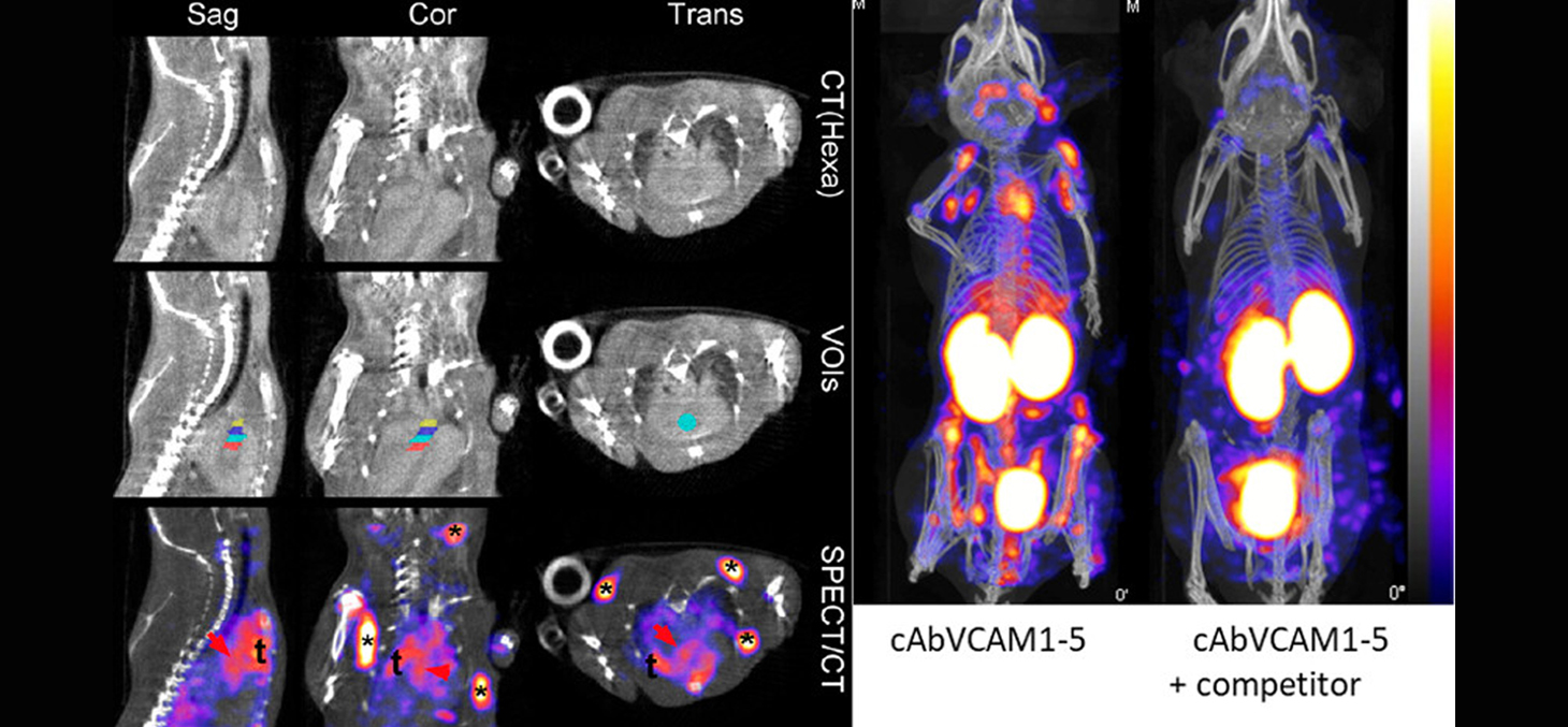
To address this, the LRB has developed 99mTc-cAbVCAM1-5, an imaging agent designed to target inflammatory processes characteristic of plaque vulnerability. This agent has been evaluated in a Phase I/IIa clinical trial in partnership with Grenoble-Alpes University Hospital (CHU). The laboratory is also developing and testing new imaging agents targeting additional biomarkers of plaque vulnerability, such as tissue factor and the urokinase receptor.
Inflammation
Molecular Imaging of Chronic Inflammation
Chronic inflammation is a key contributor to global morbidity and mortality and is considered a major public health issue due to its substantial socio-economic impact. Its early detection and monitoring are therefore critical in clinical care. Diagnosing chronic inflammation at an early, potentially reversible stage can improve treatment outcomes. It could also serve as a prognostic indicator or a tool to monitor therapeutic efficacy.
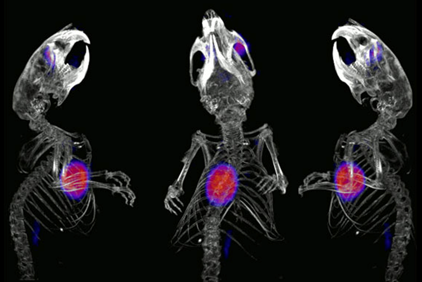
In recent years, the LRB has developed cAbVCAM1-5, an inflammation imaging agent targeting VCAM-1, a cell adhesion molecule. This agent has been validated in mice for imaging atherosclerosis and hepatic inflammation, and has already been administered to a first patient at Grenoble-Alpes University Hospital (CHU).
The current focus is on evaluating the diagnostic and prognostic potential of this imaging agent in preclinical models of chronic inflammation, including spondyloarthritis, myocardial infarction, myocarditis and neuro-inflammation.
Contact
Principal Investigators
Coronary Microvascular Dysfunction
Laurent Riou
laurent.riou univ-grenoble-alpes.fr (laurent[dot]riou[at]univ-grenoble-alpes[dot]fr)
Tel +33 (0)4 76 63 75 09
Atherosclerosis and Inflammation
Alexis Broisat
alexis.broisatinserm.fr (alexis[dot]broisat[at]inserm[dot]fr)
Tel +33 (0)4 76 63 71 02
- Share
- Share on Facebook
- Share on LinkedIn