- Share
- Share on Facebook
- Share on LinkedIn
Nuclear imaging is the gold standard for molecular imaging, utilizing SPECT (Single Photon Emission Computed Tomography) and PET (Positron Emission Tomography) cameras. SPECT cameras detect molecules labeled with gamma-emitting radionuclides, while PET cameras detect molecules labeled with positron (beta+)-emitting radionuclides.
As a non-invasive technique with very high sensitivity, nuclear imaging enables the in vivo study of physiological, biochemical, and molecular processes. Complementing morphological imaging techniques such as radiology, ultrasound, and MRI, it provides functional and metabolic insights. Widely used in clinical practice, nuclear imaging is also an extremely powerful research tool.
Equipments
The nuclear imaging laboratory is part of the Laboratoire Radiopharmaceutiques Biocliniques (LRB) and is equipped with several high-performance systems. These include a SPECT/CT scanner and a PET/MRI scanner, which can be used separately or in combination (e.g., PET/CT/MRI), depending on the needs of the study.
In addition, clinical SPECT and PET systems located in the Nuclear Medicine Department at Grenoble Alpes University Hospital (CHU) are also available for use in clinical trials.
-
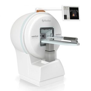
SPECT/CT Imaging: nanoScan SPECT/CT (Mediso)
Equipped with four detection heads and three sets of multipinhole collimators, this highly sensitive system enables 3D imaging of radiotracer distribution with submillimeter resolution and supports absolute quantification of biodistribution.
It is coupled with a micro-CT (X-ray) module that provides anatomical information.
-
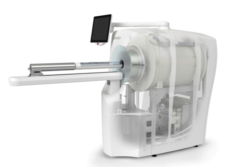
PET/MRI: nanoScan PET/MRI 3T (Mediso)
Its cryogen-free superconducting magnet is specially shielded to prevent interference with PET detection, preserving the optimal performance of both modalities.
The nanoScan PET/MRI 3T offers the highest resolution among commercially available systems, with high sensitivity. Spatial resolution reaches 0.7 mm using Tera-Tomo™ 3D iterative PET reconstruction.
The system includes a 3T MRI scanner providing both anatomical and functional imaging data.
-
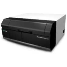
Autoradiographic Imaging
Radiotracer biodistribution can also be assessed at the tissue level with high resolution using frozen tissue sections. This is performed using a phosphor imager (Typhoon, Cytiva).
-
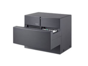
Gamma Counting
An automated gamma counter is available for absolute quantification of radioactivity in samples with high sensitivity. It is commonly used for ex vivo biodistribution studies (AMG, Hidex).
-
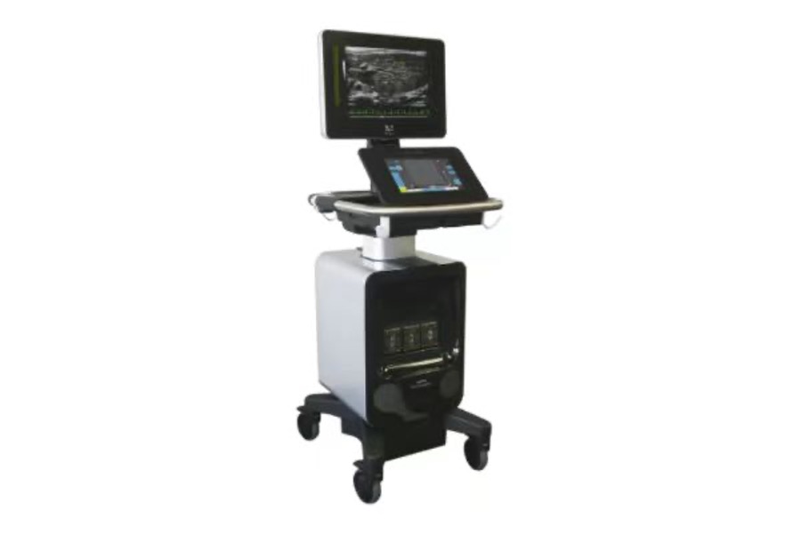
Ultasound imaging: Vevo F2 (VisualSonics - Fujifilm)
The GAIA imaging platform is equipped with a Vevo F2 ultrasound system, featuring one probe for mouse imaging and another for rat imaging.
The system supports all major acquisition modes: B-Mode, M-Mode, Doppler, Color Doppler, and Strain.
- Share
- Share on Facebook
- Share on LinkedIn